Claustrophobia and MRI Anxiety
A handout on claustrophobia and the anxiety many people feel about MRI scans: the two fears involved (suffocation and being trapped), the catastrophic thinking that drives them, and a plan combining interoceptive exposure and graded practice with enclosed spaces. For adults.
About this resource
Claustrophobia is a specific phobia of enclosed or confined spaces such as elevators, small rooms, tunnels, crowded places and, increasingly, MRI scanners. Research on claustrophobia describes two linked components: fear of restriction (being unable to move or leave) and fear of suffocation (running out of air). Both are driven by catastrophic misinterpretation of normal sensations. In a closed space the person notices warmth, a faster heartbeat and shallower breathing, interprets these as the start of suffocation or of losing control, and the surge of anxiety produces exactly the sensations that were feared. Escape or avoidance ends the episode and confirms the danger.
MRI scans are a frequent trigger and a frequent reason for referral, since a substantial number of scans are cut short or refused because of anxiety. The scanner combines enclosure, immobility, loud noise and a fixed duration, and the person cannot see out. This handout explains the claustrophobia model, offers the specific information about MRI that reduces catastrophic predictions (the tube is open at both ends, the person is monitored throughout, there is a call button, the scan is not dangerous), and describes a two-part treatment: interoceptive exposure to breathlessness and heat so that the sensations lose their threat, and graded exposure to enclosed spaces culminating in a practice session in a scanner or a scanner mock-up where available.
The professional version adds guidance on eliciting the specific feared catastrophe, on the sequence of interoceptive exercises most relevant to claustrophobia, and on liaising with imaging departments for practice sessions.
How to use it
- Identify which component dominates: fear of being unable to get out, fear of not getting enough air, or both. Ask what the client thinks would happen if they stayed in the situation for twenty minutes.
- Provide accurate MRI information from the handout and, where possible, from the imaging department. Correcting specific misconceptions (that the tube is sealed, that air is limited, that no one is watching) often reduces fear substantially.
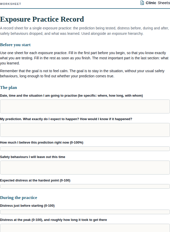
- Begin interoceptive exposure in session: breathing through a narrow straw, holding the breath, sitting in a warm small room with a coat on. Repeat until the sensations are familiar and the catastrophic belief has weakened.
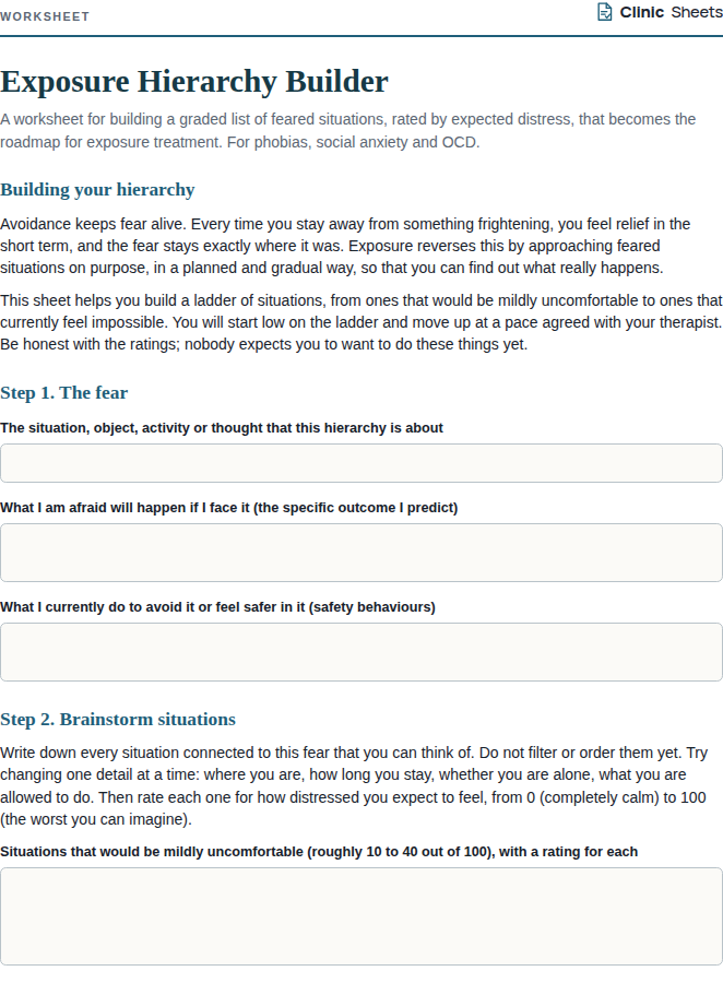
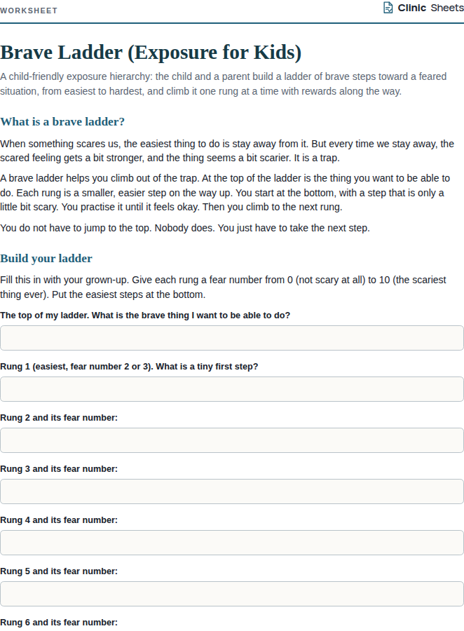
- Build a hierarchy of enclosed spaces (a closet with the door ajar, then closed; an elevator; the back seat of a small car; lying under a heavy blanket with the face partly covered; a narrow space with a lid or cover) and work through it, staying until anxiety falls.
- Where a scan is scheduled, ask the department about a practice visit or a mock scanner, and rehearse the call button, the breathing plan and the attention plan in advance.
References
- Rachman, S., & Taylor, S. (1993). Analyses of claustrophobia. Journal of Anxiety Disorders, 7(4), 281-291.
- Clark, D. M. (1986). A cognitive approach to panic. Behaviour Research and Therapy, 24(4), 461-470.
- Abramowitz, J. S., Deacon, B. J., & Whiteside, S. P. H. (2019). Exposure Therapy for Anxiety: Principles and Practice (2nd ed.). Guilford Press.